All about the skin around the eyes and “drooping” eyelids
04/04/2025

23/11/2016
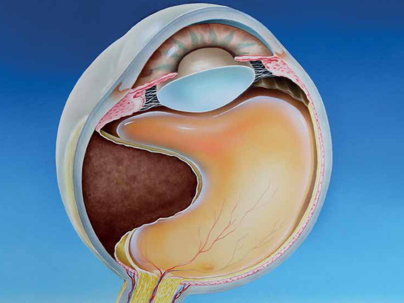
Melanoma is a tumour that appears most frequently on the skin, but it can also grow in the intraocular space from pigmented cells (melanocytes) in the middle vascular layer of the eye, the uvea, composed of iris, ciliary body and choroid. The choroid is the layer of blood vessels behind the retina that provides oxygen and other nutrients. Choroidal melanoma is the most frequent primary malignant ocular tumour in the adult and affects 6-9 people per million inhabitants per year (according to data from the United States and Nordic European countries), being more frequent in people of light skin and light eyes.
CLINICAL SIGNS AND SYMPTOMS
In many cases the patient does not present symptoms and the melanoma is diagnosed in a routine exploration. In other cases, the tumour grows in the back of the eye and gives rise to symptoms such as blurred vision or decreased visual field, whether or not accompanied by the perception of floaters (myodesopsia) or flashes (photopsies). Occasionally the growth of melanoma may produce cataracts, intraocular haemorrhages, retinal detachment or even spread to the superficial layers of the eye, making it visible as a dark spot on the sclera (white part of the eye).
DIAGNOSTIC METHODS
The diagnosis is made through a complete ocular examination that includes a fundus examination of the eye with dilatation of the pupil, specialized photographs (retinographies with and without contrast), optical coherence tomography and ocular ultrasound. In the latter, the melanomas have very typical characteristics that confirm the diagnosis and also measure the tumour to assess its size and degree of activity or growth over time. With these tests and in expert hands, the diagnostic accuracy exceeds 95%. Since melanoma is a type of cancer with metastatic spread, once the ocular diagnosis has been made it is imperative to carry out a study and follow-up of tumour extension, with the collaboration of a specialist in medical oncology, to assess the possible compromise of the periocular tissues and other organs (liver and bones, fundamentally).
TREATMENT
Treatment options for eradicating the tumour are multiple and highly specialized. For 15 years, the Ocular Oncology Unit of the Barraquer Ophthalmology Centre, led by Prof. Rafael I. Barraquer, has been offering a comprehensive and pioneering service in Spain for the diagnosis and treatment of ocular tumours. In the case of melanoma, depending on the size and location of the tumour, we use radiotherapy (brachytherapy with radioactive isotopes or different modalities of external radiotherapy) or advanced techniques of ocular microsurgery for resection of the tumour. The enucleation of the eyeball continues to be a necessary alternative only in very advanced cases, in which the melanoma is of great volume and can no longer be treated with conservative methods.