All about the skin around the eyes and “drooping” eyelids
04/04/2025
19/11/2021
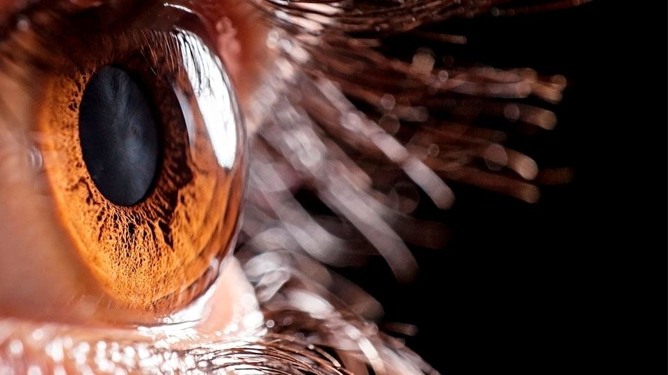
A corneal examination can show the presence of human diseases. Inspection by an experienced professional using a simple instrument (slit lamp) can give a more accurate diagnosis than most modern imaging tests. The ophthalmologist can diagnose the disease using the examination.
There are infinite examples of a clinical diagnosis from a corneal examination:
► The Herpes Zoster virus is associated with punctate or dendritic keratitis, lymph nodes, opacities and hypoesthesia. The Epstein Barr virus causes stromal keratitis and subepithelial infiltrates. HIV is associated with dry eye.
► Tuberculosis can manifest as interstitial keratitis through a reaction to tuberculins. Spread by ticks, Lyme disease will cause a corneal lesion from secondary exposure to facial paralysis. Chlamydia causes trachoma, the number one preventable cause of blindness throughout the world. Amebiasis is associated with poor contact lens hygiene. It affects healthy patients.
► Sjögren’s syndrome is dry eye disease. It is associated with scleroderma, lupus, rheumatoid arthritis, psoriatic arthritis and juvenile arthritis. Cogan’s syndrome is associated with deafness, vertigo and bilateral interstitial keratitis. Polyarteritis nodosa shows an ulcered ring in the corneal margin. Sarcoidosis: dry eye and interstitial keratitis. Conjunctival pemphigoid causes exposure keratopathy with epithelial defects.
► Hemochromatosis is associated with limbal iron deposits and pseudoverticillata cornea. Cystinosis produces intracellular accumulation of cystine crystals: they are deposited in the central and peripheral epithelium. Ehlers-Danlos syndrome is associated with corneal ectasia (keratoconus and keratoglobus). 10% of patients with Down’s syndrome present with keratoconus.
► Multiple myeloma accumulates proteins in the epithelium and stroma. Leukaemia can cause direct infiltration of the cornea through the disease's cells.
► There is involvement due to chronic lack of irrigation in the arteriosclerotic disease: neuroparalytic keratitis.
► Diabetes mellitus leads to a loss of corneal sensitivity and poor epithelial healing: punctate keratitis, recurring erosions and neurotrophic ulcers. Gout: uric dystrophy, limbal subepithelial opacities, ulcers and neovascularisation.
► A shortage of vitamin A may cause nyctalopia (night blindness), xerophthalmia, punctate keratitis, superficial keratinised opacities. Hypervitaminosis D can cause corneal califications such as band keratopathy.
► The use of amiodarone causes yellowish deposits in the epithelium and cornea verticillata. Anti-malarials, greyish-white dots on the inferior cornea, along the Bowman's membrane.
In the consulting room we can use complementary tests to confirm the suspected diagnosis. This is one more reason to stress the importance of an eye check-up at the same time as a check-up on our general health.