All about the skin around the eyes and “drooping” eyelids
04/04/2025

22/01/2021
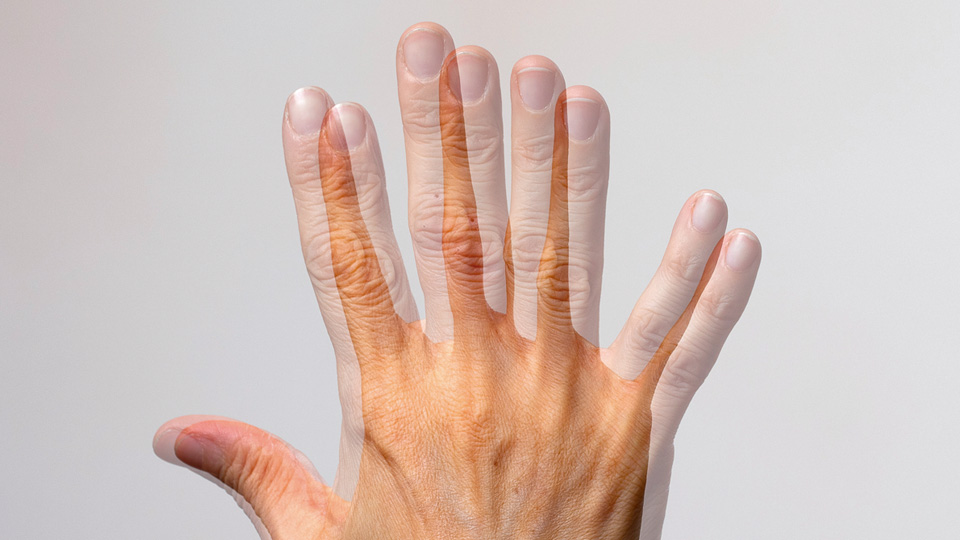
Diplopia or double vision is the perception of two images of one single object. It happens because each eye perceives the object in a different point in the space, and the brain interprets it as if there were two objects.
Types and causes
Monocular diplopia: The double image is perceived with just one eye open. It is due to structural alterations to the eyeball, the most common being:
Crystalline lens alterations: cataracts, crystalline lens subluxation.
Corneal abnormalities: keratoconus, corneal scarring or opacity.
Non-corrected refractive errors: astigmatism.
Macular pathologies: epiretinal membrane.
Binocular diplopia: This is most common type. It appears with both eyes open and disappears upon occluding one of them. It is caused by a lack of parallelism in both eyes because of an alteration to the oculomotor system. Although there are many diseases that may affect the alignment of the eyes, the most common are:
Childhood strabismus compensated in adulthood
Oculomotor nerve palsy
Neurological diseases (myasthenia gravis)
Thyroid diseases
Brain tumours
Cranial and orbital trauma
Eye surgery
High myopia
When the diplopia is binocular, the patient’s head occasionally adopts an abnormal position (torticollis) to compensate the double vision.
Diagnosis
At the consultation, the ophthalmologist will ask for the patient’s medical history to obtain details on the way the diplopia appeared, its duration, if its constant or intermittent, if it is accompanied by other symptoms and if the patient suffers associated risk factors. In addition, they will undertake an examination of the anterior and posterior segment of the eye, a pupil reaction test and an ocular motility exam to determine the degree of deviation and the muscles causing it. In some cases, complementary tests will be required (analysis, neuroradiological study) to rule out a systemic compromise.
Treatment
The prognosis will depend on the cause of the diplopia. Once diagnosed and treated, if the binocular diplopia persists, there are three therapeutic options.
Dr. Idoia Rodríguez Maiztegui, ophthalmologist at the Barraquer Ophthalmology Center