All about the skin around the eyes and “drooping” eyelids
04/04/2025

03/01/2023
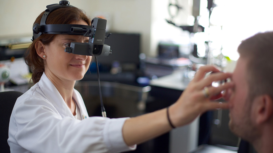
The examination of the fundus, technically known as ophthalmoscopy, is one of the most common exams in ophthalmological check-ups. It is a simple and painless test that give us complete information about the main structures of the back of the eyeball: optic nerve, macula, vascular arcades, peripheral retina and choroid. The results obtained help us diagnose and monitor different eye pathologies and, eventually, decide the best treatment for them.
Ophthalmologists perform the examination using an instrument called an ophthalmoscope, a device that allows for visualization of the back of the eye through a light source and a lens system. In most cases it is necessary to previously dilate the pupil with the application of eye drops, as the access is through the pupil. For this reason, we recommend to bring someone with you since after the examination is carried out the pupils will continue to be dilated and the vision will be blurred for a few hours.
Frequent pathologies such as age-related macular degeneration, myopia or diabetic retinopathy, among others, can be diagnosed with this innocuous test. For this reason, at the Barraquer Ophthalmology Centre, we recommend a periodic evaluation of the fundus to rule out these diseases and maintain good eye health.
Dr. Sònia Viver, ophthalmologist at the Barraquer Ophthalmology Centre